The Nuclear Medicine and Radiation Biology (NMRB) research group at UiT The Arctic University of Norway conducts interdisciplinary research at the intersection of nuclear medicine, molecular imaging, radiopharmaceutical sciences, and radiation biology. In close collaboration with the University Hospital of North Norway (UNN), we combine advanced imaging technologies, radiopharmaceutical development, and experimental research to improve our understanding of disease and advance new diagnostic and therapeutic strategies.
Our research focuses on the development of novel radiopharmaceuticals for imaging and targeted radionuclide therapy, the application of PET and other molecular imaging techniques, radiation-based treatments, and personalized medicine. By integrating preclinical investigations, clinical imaging, and computational approaches, including machine learning, we aim to identify new biomarkers, investigate disease mechanisms, and improve the assessment and treatment of cancer and other diseases.
Supported by advanced imaging and radiopharmaceutical production facilities in Tromsø, NMRB provides a collaborative environment for researchers, clinicians, and students. Our goal is to translate scientific discoveries into innovative imaging methods and more effective, individualized healthcare through national and international research partnerships.
PET-core is a preclinical research facility jointly operated by UiT The Arctic University of Norway and the University Hospital of North Norway (UNN). The facility provides specialized equipment, technical support, and scientific expertise for researchers investigating disease processes, developing new radiopharmaceuticals, and evaluating innovative approaches to molecular imaging and radionuclide therapy.
By enabling detailed studies in experimental models, PET-core helps researchers investigate how imaging agents and therapeutic compounds behave in biological systems. The facility supports research from early-stage radiotracer development and experimental validation to the assessment of potential diagnostic and therapeutic applications.
Booking and access to services
Researchers who wish to use PET-core facilities and services can access the booking system through the UiT PPMS platform. The platform provides access to the facility's service booking system and related information. Users should consult the platform for available services and booking procedures.
Facilities and Research Technologies
PET-core brings together complementary imaging systems and laboratory resources that allow researchers to study biological processes, characterize radiotracers, and investigate the effects of targeted radiation. Available infrastructure includes:
Small-animal imaging: Integrated PET, SPECT, and CT systems for investigating tracer distribution, biological function, and disease-related changes.
Combined PET/MRI: Simultaneous acquisition of molecular and anatomical information for multimodal studies.
Image-guided irradiation: A stereotactic irradiation system with optical and CT guidance for precise delivery of radiation in experimental models.
Real-time autoradiography: A platform for studying the spatial distribution of radioactive compounds in biological samples.
Cell laboratory: Laboratory facilities for cellular experiments supporting preclinical research.
Radiotracer characterization: Instrumentation for evaluating and validating novel radiotracers and supporting their development for research applications.
Together, these resources enable researchers to design experiments that combine molecular imaging, laboratory analyses, and radiation-based interventions. The facility supports studies in oncology, molecular imaging, radiobiology, and the development of targeted diagnostic and therapeutic strategies.
From Experimental Research to Clinical Applications
An important objective of PET-core is to help bridge the gap between experimental discoveries and their potential use in clinical medicine. Preclinical investigations allow researchers to assess the performance of new imaging agents, explore their biological distribution, investigate treatment responses, and generate evidence needed to guide further development.
PETcore's infrastructure supports research into:
New radiotracers for disease detection and characterization
Molecular imaging biomarkers and quantitative imaging methods
Radiopharmaceuticals for targeted radionuclide therapy
Biological responses to irradiation and experimental treatments
Theranostic approaches that combine diagnostic imaging with targeted therapy
Imaging-based assessment of treatment efficacy and disease progression
The facility also provides a setting for multidisciplinary projects combining experimental biology, radiochemistry, image analysis, engineering, and computational methods. This approach supports the development of more informative preclinical studies and helps researchers evaluate promising technologies before progression to clinical investigation.
Research, Training and Collaboration
PETcore supports research projects at UiT, UNN, and collaborating institutions. Its infrastructure is available as a resource for scientific projects requiring specialized preclinical imaging, experimental irradiation, or radiotracer evaluation, subject to project requirements and facility arrangements.
The facility also contributes to research education and professional development. Students, doctoral researchers, clinicians, and other scientific personnel can develop practical knowledge of preclinical imaging methods and their applications in biomedical research.
PETcore is connected to the European Infrastructure for Translational Medicine (EATRIS), providing a link to a wider European research environment focused on translating biomedical discoveries into medical applications.
Collaboration opportunities include joint research projects, development and evaluation of radiopharmaceuticals, validation of imaging biomarkers, experimental studies of targeted therapies, and projects incorporating image analysis and machine learning.
Researchers and external partners interested in using PETcore's facilities are encouraged to contact the team to discuss their scientific objectives, technical requirements, and potential collaborations.
The PET-core Team
PETcore brings together expertise in preclinical PET imaging, research infrastructure, engineering, and computational methods. The team supports the planning and execution of research activities and the effective use of the facility's technologies.
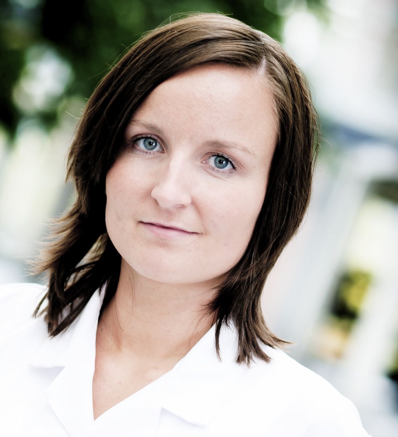
Montserrat Martin-Armas
Head of Preclinical PET-core Facility and Senior Researcher
Montserrat Martin-Armas leads PET-core and contributes to its research activities and scientific development.
For enquiries about PET-core's facilities, research collaborations, technical capabilities, or access to preclinical imaging infrastructure, please contact the facility team:petcore@uit.no
The Chemistry Facilities at the PET Imaging Center in Tromsø provide advanced infrastructure for the production, development, and characterization of radiopharmaceuticals for molecular imaging and targeted radionuclide therapy. The facilities integrate cyclotron-based radionuclide production, radiochemistry, radiosynthesis, and analytical quality control, supporting research from the development of novel radioactive tracers to their evaluation for potential clinical applications.
Equipped with a dedicated cyclotron, specialized radiosynthesis laboratories, advanced radioanalytical instrumentation, and GMP-compliant radiopharmaceutical production facilities, the centre supports the development of new radiotracers for PET imaging and theranostics. These capabilities enable interdisciplinary collaboration between chemists, radiochemists, physicists, biologists, and clinicians, strengthening translational research and advancing precision medicine.
Cyclotron
The cyclotron is a 16.5/8.4 MeV PETtrace 800-series cyclotron with dual extraction. Targets for18F,11C (CO2and CH4),13N, and15O are available, generating a wide range of possibilities in terms of radiolabelling. In addition, the solid target unit Alceo (Comecer) will make the production of metal radionuclides such as 64Cu 89Zr and 45Ti possible. Read more
Radiopharmaceutical Development Unit
Radiosynthesis lab
2 MIP shielded cells (Comecer)
TRACERlab FxC (11CO2 and 11CH3I/TfO)
TRACERlab FxN (18F)
Safety bench for manual radiosynthesis intended for 99mTc
Cold precursor synthesis at the Dept. of Chemistry, UiT
Radioanalysis lab
High-resolution UHPLC-QToF-MS including DAD detection (6530 model) and metabolomics analysis software (Agilent)
Analytical HPLC/UHPLC fitted with DAD and radioflow beta coincidence detection (Agilent and PosiRam detector from Lablogic) for pharmacokinetic studies (together with our HR-QtoF MS)
Head-Space-GC-FID for residual solvent determination (Agilent)
High-purity Germanium MCA (gamma spectrometry)
RadioTLC (Lablogic)
Dose calibrator
Rotavapor, centrivap, refrigerated centrifuge, osmometer, etc.
GMP Radiopharmaceutical Unit
In the GMP radiopharmaceutical production area, eight BSS series shielded cells and two Talia dispensing shielded cells are available for further product development into clinical application.
The Clinical Facility at the PET Imaging Center in Tromsø provides advanced molecular imaging services for patient care, clinical research, and the development of innovative diagnostic approaches. Located at the University Hospital of North Norway (UNN), the facility integrates state-of-the-art PET/CT, PET/MRI, and SPECT/CT technologies to support the diagnosis, characterization, and monitoring of a wide range of diseases.
A major addition to the facility is the Siemens Biograph Vision Quadra full-body PET/CT scanner, an advanced imaging system that expands the possibilities for whole-body molecular imaging. Its extended axial field of view enables the acquisition of comprehensive PET data with high sensitivity, supporting advanced imaging protocols, quantitative analyses, and research into disease processes throughout the body. This technology creates new opportunities for clinical research, novel radiopharmaceutical evaluation, and the development of personalized diagnostic and therapeutic strategies.
Clinical imaging at the facility supports research and patient care across oncology, neurological disorders, cardiovascular medicine, and other areas where molecular information can improve disease characterization and treatment assessment. By visualizing biological and metabolic processes in vivo, PET imaging provides valuable insights into disease mechanisms, treatment response, and disease progression.
Through close collaboration between UNN and UiT The Arctic University of Norway, the Clinical Facility connects clinical expertise, advanced imaging technology, and translational research to advance nuclear medicine, precision diagnostics, and the development of new approaches to patient care.
THERACAN – Targeted Radionuclide Treatment of Cancer
Project period: through December 2030
THERACAN aims to develop more targeted treatments for aggressive cancers using radioactive substances. The project combines expertise at UiT and the University Hospital of North Norway (UNN) to advance radionuclide-based treatment strategies and support the development of more individualized cancer care.
By connecting radiopharmaceutical research with nuclear medicine and translational studies, THERACAN seeks to improve how radioactive compounds can be used to target cancer while supporting the development of new therapeutic approaches.
Funding: Tromsø Research Foundation, Trond Mohn Foundation, and the Norwegian Cancer Society.
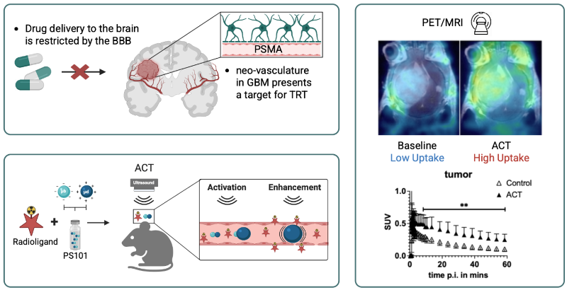
NORTHCARE (Novel theranostic application of radiotherapeutics for personalized cancer treatment using focused ultrasound with Positron Emission Tomography) is a research project funded by the Tromsø Research Foundation and focused on developing advanced theranostic strategies for personalized cancer treatment. The project combines diagnostic imaging and targeted radiotherapy in a single platform, aiming to improve both treatment precision and outcome monitoring. By integrating focused ultrasound with PET and SPECT imaging, NORTHCARE seeks to enhance the delivery and effectiveness of radiolabeled therapeutic agents while allowing real-time assessment of tuuptake and response.
The 180°N consortium brings together universities and university hospitals in Tromsø, Trondheim, and Bergen, with additional participation from Stavanger. Its research focuses on improving cancer diagnostics and treatment, developing new approaches for neurological disorders, and enabling more precise patient stratification and personalized medicine.
The Tromsø research activities emphasize preclinical testing and evaluation of innovative imaging and therapeutic approaches. The programme includes four major areas: immuno-PET for lung cancer, targeted radionuclide therapy and imaging for glioblastoma, phagocyte-targeted approaches in breast cancer, and machine-learning methods for oncological PET imaging.
NMRB principal investigator: Rune Sundset. Funding: Tromsø Research Foundation and Trond Mohn Foundation. The listed programme period is 2019–2025; confirm whether follow-up activities continue before labelling it ongoing.
SECURE – Novel Marine Resources for Food Security and Food Safety
Interdisciplinary research in nutrition and cardiovascular health
SECURE investigates how novel marine resources from the Arctic, including microalgae, Calanus, and sea cucumber, may contribute to sustainable food systems, food safety, and cardiometabolic health. The project brings together expertise in marine resources, nutrition, microbiology, cardiovascular research, and regulatory science.
NMRB contributes through preclinical radionuclide imaging research investigating atherosclerosis. PET imaging offers a non-invasive method for studying atherosclerotic plaque development and characterizing lesions, helping researchers explore relationships between diet, biological processes, and cardiovascular disease.
Funding: UiT Thematic Research Areas. Imaging work package leaders: Rune Sundset and Karl-Erik Eilertsen.
BrainSTORM – PET in Targeted Brain Theranostics and Education
Project period: 2019–2022
Brain theranostics holds tremendous promise for treatment of brain disease, involving design and development of molecular probes for imaging, monitoring and therapy. However, significant challenges remain, such as blood-brain barrier (BBB) permeability, targeted and controlled delivery to specific brain regions as well as early disease diagnosis. The BrainSTORM project aims to address these challenges via multidisciplinary approaches to personalized precision medicine in the areas of Alzheimer ́s disease, depression and brain cancer. The Positron Emission Tomography (PET) Imaging Center in Tromsø opened in 2018 as a state-of-the-art facility for clinical and preclinical PET/CT, PET/MR and SPECT/CT imaging as well as base for the generation of radioactive isotopes and molecules. The co-localization with established UiT research environments in medicine, chemistry, physics, pharmaceutical sciences, biology and science education enable us to develop cutting-edge brain theranostics for personalized precision medicine and an integrated unique educational program. Objectives of the BrainSTORM project include:
Develop PET/SPECT-radiotracers and nanoparticle delivery systems selectively targeting brain receptors central to Alzheimer ́s disease, depression and cancer.
Apply radiotracer systems to cutting-edge theranostics (therapy and diagnostics) and basic brain function studies on animal models.
Advance secondary and tertiary education within interdisciplinary science in health technology and its use in research, treatment and diagnosis of brain diseases.
Explore the latest scientific publications from the Nuclear Medicine and Radiation Biology (NMRB) research group and our collaborators. Our research advances molecular imaging, radiopharmaceutical development, nuclear medicine, and radiation biology, contributing to new diagnostic and therapeutic approaches.
PETcore supports master's and PhD students conducting research in preclinical imaging, molecular imaging, radiopharmaceutical development, quantitative PET, machine learning, and translational medicine. These projects provide opportunities to work with advanced imaging technologies and collaborate with researchers and engineers at UiT The Arctic University of Norway and the University Hospital of North Norway (UNN).
This section presents current student projects, including research topics, student names, supervisors, and expected completion dates where available.
PhD projects
Elin Kile Virtual Biopsy of Prostate Cancer Using PET Imaging and Artificial Intelligence
PhD project in Health Sciences, listed in 2023.
Supervisors: Samuel Kuttner (main supervisor) and Rune Sundset (co-supervisor).
This project investigates the use of PET imaging and artificial intelligence to support the characterization of prostate cancer.
This section highlights master's theses and doctoral dissertations completed through research projects associated with PETcore and its collaborating research groups. The projects reflect the facility's contributions to molecular imaging, nuclear medicine, radiopharmaceutical development, image analysis, and translational biomedical research.
Each entry includes the student's name, thesis title, degree, year of completion, supervisors, and a link to the full thesis where available.
PhD theses
Elena Markova
Thesis title:Eat, Deliver, Destroy: How Liposomes and Phagocytes Team Up
Research area: Nanomedicine, drug delivery, and cancer research.
Elena Markova's doctoral research investigated interactions between liposomes and phagocytes, exploring approaches to improve the delivery of therapeutic compounds and support targeted cancer treatment.
Thesis title:On-site Cyclotron Produced Cu-67 for Translational Radiotheranostics
Research area: Radiopharmaceutical development, copper-67 production, and theranostics.
Ursula Søndergaard's doctoral research focused on developing and validating methods for producing copper-67 using a biomedical cyclotron and evaluating its potential for translational radiotheranostics. The work supports research into more accessible production of therapeutic radionuclides and targeted cancer treatment.
Lene Brosvik Radiosynthesis and Binding Evaluation of Novel CDK7-Targeting C-11 Radioligand for Glioblastoma Nuclear Imaging
Programme: Health Sciences Year listed: 2024 Supervisors: Angel Moldes-Anaya (main supervisor) and Mathias Kranz (co-supervisor).
This project focused on the development and evaluation of a carbon-11-labelled radioligand targeting CDK7, with potential applications in glioblastoma imaging.
Daniel Sakarli PET Reconstruction-Dependent Image Quality and Quantification
Programme: Medical Engineering, Friedrich-Alexander University, Erlangen, Germany. Supervisor: Samuel Kuttner.
This project investigated how PET image reconstruction methods influence image quality and quantitative measurements.
Rune Sundset at Trond Mohn Annual Celebration at Aula in Bergen, Norway on December 01, 2023.
Photo credit Melanie Burford
Fredrik Aspheim, Luigi Luppino, and Samuel Kuttner were co-authors of work that were awarded for best presentation at the Artificial Intelligence and Informatics in Nuclear Medicine Symposium Symposium (AINM) 2023
Assoc. Prof. Tom Christian Holm Adamsen , Rune Gildsig, Irene Skaar (PhD), Ole Heine Kvernenes (PhD), Karl Fredrik Strømsheim Alnes (PhD), Haukeland University Hospital