Faculty staff
Fred GodtliebsenProfessor

Michael KampffmeyerProfessor
Michael Kampffmeyer – Professor
Contact:
E-mail: michael.c.kampffmeyer@uit.no
Work phone: +4777625264

Puneet SharmaAssociate Professor
Puneet Sharma – Associate Professor
Contact:
E-mail: puneet.sharma@uit.no
Work phone: +4777660391
Private phone: 45063429

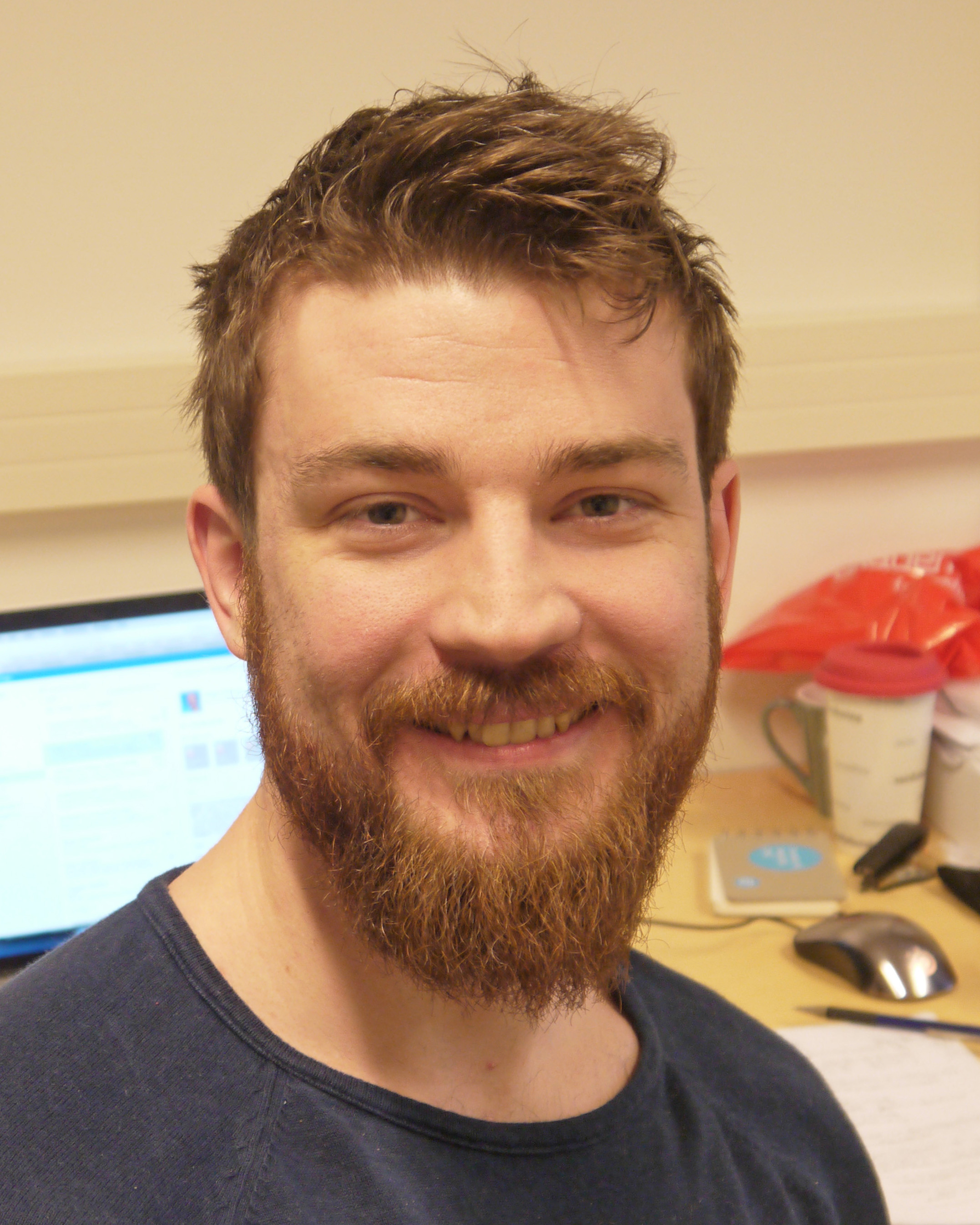
Kristoffer WickstrømAssociate Professor (Group leader)
Kristoffer Wickstrøm – Associate Professor
Contact:
E-mail: kristoffer.k.wickstrom@uit.no
Work phone: +4777623216

Robert JenssenProfessor

Elisabeth WetzerAssociate Professor
Elisabeth Wetzer – Associate Professor
Contact:
E-mail: elisabeth.wetzer@uit.no
Work phone: +4777644874

Qing LiuAssociate Professor

Georgios LeontidisProfessor

Veronica LachiAssociate Professor

Benjamin RicaudAssociate Professor
Benjamin Ricaud – Associate Professor
Contact:
E-mail: benjamin.ricaud@uit.no
Work phone: +4777625247